
As a Physiotherapist working in Women’s Health, I talk about the “Pelvic Floor” with most of the clients I see. However, many of the ladies I treat are unsure about where their pelvic floor is and what it does.
But if a muscle can’t be seen or visualised, how can we expect women to connect with it well when they are told to exercise it?
This aim of this blog post is to provide some detail about the anatomy of the Pelvic Floor, it’s function and importance, particularly in relation to its role in Women’s pelvic health conditions.
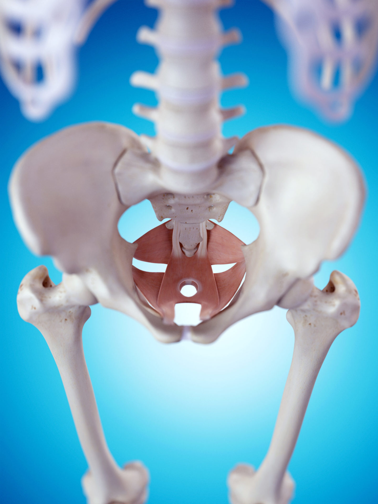
The Pelvic floor is a large, layered sling of muscle which is situated at the base of the pelvis or ‘undercarriage’. It connects to the front, back and side walls of the pelvic bones, where the two pelvic bones meet. It helps to provide support and stability to the Lumbopelvic region and to the internal structures within the pelvis.
The pelvic floor muscles can be divided into three layers. The deepest layer is called the Pelvic Diaphragm. It consists of the Levator Ani, which are a group of muscles which play an important role in supporting the pelvic organs and in maintaining continence. This layer also includes some important hip and bottom muscles, which help to provide support and movement to this area of the body.

The middle layer of the pelvic floor is called the Urogenital diaphragm. This layer consists of several muscles which act as sphincters to support the walls of the urethra and vagina and help to keep them closed.
The most superficial layer is called the Urogenital Triangle. It can be felt externally, around the sides of the vulva and the top of the groin region. These superficial muscles play an important role in sexual function and can contribute to problems with intercourse and pelvic pain conditions. This layer also consists of the external anal sphincter which helps to keep the anus closed and is therefore important in maintaining fecal continence.

Other important pelvic structures
Within the pelvis, the uterus, bladder and bowel are well supported in their relative positions by the pelvic floor muscles, but also by a dense network of soft tissues or ‘fascia’, also termed the Endopelvic fascia. This soft tissue network connects with the muscles and bones within the pelvis
The Pudendal nerve is one of the most important nerves found within the pelvis and is important in providing normal sensation and muscular activation. It may be damaged during vaginal birth and this can contribute to pelvic health problems, such as incontinence.
Common symptoms that may indicate pelvic floor dysfunction
So now we know where the pelvic floor muscles are and what they do, it is clearer as to how symptoms can develop when they become dysfunctional or damaged. These symptoms may include;
Pain during sex
Constipation
Problems with urination or defecation
Urinary or fecal incontinence
Sensations of pressure/dragging/aching and Pelvic organ Prolapse
Pelvic and vulval pain
Bladder urgency and frequency
Exercising the pelvic floor is therefore an important part of the maintenance of female pelvic health throughout life, and is particularly important during pregnancy, during the postnatal period and in the peri and post-menopausal periods. Pelvic floor exercise is also an important component in the rehabilitation of pelvic health problems such as prolapse or incontinence.
But how should you be exercising your pelvic floor? Our follow-on blog “How to exercise your Pelvic Floor” details how you should be exercising these muscles appropriately, to suit your individual need and ability.
If you have any specific questions or concerns about your pelvic health, please contact us directly.
